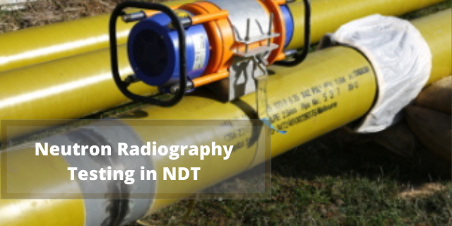
22 Jan Neutron Radiographic Testing in NDT
What is Neutron Radiography Testing
Neutron radiography technology is also called Neutron Imaging, Neutron Tomography or else N-Ray Radiography. This is a powerful NDT method because of the neutron’s properties. It allows imaging of materials which are hydrogenous like oil, plastic, water, rubber, explosives etc, which should be within the components which are made up of brass, steel, aluminium and nickel. Even though the mechanisms of N-ray and X-ray are similar , the penetration depth of different materials in each type of radiation provides a new perspective for the N-ray method.
This is a powerful method since it can view through many complex materials , corrosion products and identify the flaws and defects which cannot be notified otherwise, such as voids and cracks in energetic materials, structural weakness in composites, ceramic remnants in blades of turbines and 3D printed components. Regardless of the development of this method before decades, it has not yet fully acquired by the NDT community since it is having over-reliance on the facilities of ageing nuclear reactors.
How it works
Neutron imaging consists of a stream of neutron radiation which is used to visualise an object’s internal structure.As mentioned earlier X-rays and N-rays are similar in mechanisms but X-rays can go through light materials much more easily than dense materials. As the number of electrons in the elements and the energy consumed is directly proportional , in this case the energy required to pass through the material will be more for elements having more number of electrons, also heavier atoms will have much bigger and greater populated electron clouds which make them more opaque. N-rays mainly interact with the atomic nucleus and ignore dense electron clouds.It’s difficult for neutron radiation to pass through lighter materials because of the insufficient space between the atomic nuclei for the radiation to penetrate through it. So the neutron radiography is mainly performed on dense materials more easily than the lighter materials.
The different sources of neutrons for radiography mainly are:-
- Atomic reactors
- Radioisotopes (preferably 252 Californium)
- Particle accelerators
Imaging Techniques
There are mainly five types of imaging techniques such as:-
- Film Based Imaging
- Dynamic System Imaging
- Cold Neutron Radiography
- Fast Neutron Radiography
- Computerized Tomography
- Film Based Imaging
Film based imaging is widely used by most of the neutron radiography applications. Basically there are two film-based image recording methods, let’s discuss it in detail below:-
-Direct Exposure Technique:
In direct exposure technique, a converter foil like gadolinium is used and is placed in direct contact with the photographic film which is placed behind the object into the beam of neutrons.
The converter foil will absorb the neutrons and emit gamma-radiation and this will internally get converted into electrons.These electrons expose the emulsion facing the converter i.e gadolinium foil. This technique involves overlaying the auto-radio graphic second image and blurring of the image , so it is not applicable for radioactive objects.
-Transfer Technique:
In transfer technique, a metal foil for e.g. indium, dysprosium, gold etc is used in the place of an image recorder. Radioactivity is formed and an activation image is created in the foil after neutron absorption, which is directly transferred into the laboratory to a photographic film by using the 10 decay radiation(β radiation).
In this method, the process of decaying starts during the time of neutron exposure in the beam and which in turn loses some of the emitted radiation, which makes this technique slower than the direct exposure technique. But as an advantage this method can be used in the case of gamma ray fields since the foil is insensitive to gamma rays.
-Track-Etch Detector:
In Track-Etch Detector technique, the converter screen emits the α particles in response to incoming neutrons.An image is produced by the α particles in the nitrocellulose film by formation of small defects or tracks, which are made visible by using an etching process. In thermal neutron radiography, 11 Nitrocellulose film is extensively used, especially in N-ray radiography of radioactive objects.
-Imaging Plates (IP):
Imaging Plates (IP) techniques contain Gd as a neutron absorb-er and BaFBr:Eu 2+ as the agent (provides the photo luminescence). An imaging plate scanner is used for extracting the latent image information in the form of digitised data file by de-excitation of laser signal from the plates.
- Dynamic System Imaging
In the dynamic system method, the radiation strikes on a phosphor screen after penetrating through an object. This method is also referred to as radioscopy imaging, The image produced on the screen is later intensified and viewed using a video camera.
- Cold Neutron Radiography
In this method, the Cold neutrons are produced using a spinning chopper by filtering the low-energy end of the thermal neutron spectrum , or by using a moderator cooling with liquid nitrogen or by using cryogenic cooling. While cold neutrons (~ mill eV) penetrate materials less than the thermal neutrons, the differential attenuation between two materials may become higher, depending on the materials and precise energies involved which result in considerable improvement in contrast between two materials. The core of CNRF( Cold Neutron Research Facility ) is a cold neutron moderator which consists of a spherical shell of liquid hydrogen with a temperature of approx 20 K, placed adjacent to the reactor core.
- Fast Neutron Radiography
In Fast neutron Radiography, there is greater penetration power when compared to thermal neutrons and also present availability of mobile or any portable fast neutron sources. The variation in attenuation is very little among the elements when energies of neutrons are in the MeV range. Most materials fall in the range of 1-10 barn range (cross sections) for neutrons with MeV energies. The penetration of fast neutrons through most of the materials is quite better So its is possible to image thick objects where sufficient density changes are being attempted. cross-section in the mill barns region.
- Computerized Tomography
In transmission tomography technique, an object is positioned into a fine beam and the attenuation is calculated with a line camera from different angles. Either the source or the object and camera must be rotated in order to get a multi angle set of projections. The image of all projections is termed as sinogram since the shadow lines show sinusoidal shape. It contains all the information regarding the absorption density in a single cut.
- Internal flaws inspection such as voids, foreign materials, cracks, density variations, inclusions, bubbles and misalignment
- Testing(Reliability) of detonators in explosive devices
- In Medical field, the detection of tumours and use of doping materials to increase contrast
- Radioactive objects inspections
- Plastic coating – insulation in coaxial cable inspection and also block insulation in high voltage electrical connection Inspection
- Hydrogenous foreign substances testing in sealed units.
- Plating Inspection like cadmium plating
- Inspection of bonding flaws in adhesives.
- Gasket inspection
- Detection of artefacts uncovered through archaeological digs
- Inspection of air-cooled jet engine turbine blades
- High-reliability explosives Inspection for checking the presence of receivers and transmitters
Enroll for 1 month preparatory classes for Neutron Radiography Testing Certification
Gamma NDT academy offers NDT preparatory classes for candidates who intend to appear for the ASNT Level 2 and 3 Neutron Radiography Testing certification exam.
We will cover all the syllabus under ASNT for Neutron Radiography Testing certification exam and conduct mock tests to prepare you for the exam. We will also share study materials and sample questions which you need to revise. We have a 100% success rate for NDT certification courses.
Gamma NDT academy training institute is located in Kerala, India. We provide online classes for candidates who cannot attend our training sessions in-person.


Sorry, the comment form is closed at this time.